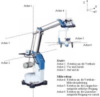
Microscope-based Assistance System
Project Description
A lot of diagnose and treatment systems, all requiring much space, are used in today's operating theaters. In this context, the microscope is a very important device. At the moment, all these instruments have specified application fields, which can't be combined.
Motorization
In cooperation with the companies Möller-Wedel and IBG Technology the HI-R1000 microscope produced by Möller-Wedel was motorised completely to support the surgeon. Now the positioning can be carried out with a remote control which is fastened to one of the instruments without interrupting the work flow of the surgeon. The remote control offers different modes with which often appearing movements can be done automatically. This encloses the pivotation around the current focus point to look at objects from another point of view, movements of the objective parallel to the current image plane to have a look at objects slightly out of sight, as well as corrections of the working distance and the zoom.
The remote control communicates with an external PC using Bluetooth. The PC determines the coordinates of the target position and calculates the positions of all joints. The corresponding parameters are sent to the microscope using a serial interface.
However, the microscope can also be positioned manually. This makes sense for movements with large amplitude which have to be carried out fast but inexactly. Moreover, this offers another security aspect, because by the manual control all motor movements are interrupted immediately.
Presenting additional information
It is also possible to display preoperative data in the actual microscope view with a special mirror system. The MRT- and X-ray-images shall be presented at the correct position and the origional size. On top of this, tissue and vessel structures will be calculated from these images and will help the surgeon to navigate.
Integration of Optical coherence tomography
Optical coherence tomography (OCT) is a relatively new method, which can be used for diagnosis by the surgeon. An OCT system will be integrated in the motorized microscope and can be positioned automatically.
Publications
2010
An experimental comparison of control devices for automatic movements of a surgical microscope, Geneva, Switzerland , 2010. pp. 311-312.
Motorization of a Surgical Microscope for intra-operative navigation and intuitive control, International Journal of Medical Robotics and Computer Assisted Surgery , vol. 6, no. 3, pp. 269-280, 2010.
| DOI: | 10.1002/rcs.314 |
| File: | rcs.314 |
2008
3D Simulation of a motorized operation microscope, Venice, Italy: Springer-Verlag Berlin Heidelberg, 2008. pp. 258-369.
Intraoperative Fernsteuerung eines Operationsmikroskopes, Bartz, Dirk and Bohn, S. and Hoffmann, J., Eds. Leipzig, Deutschland; Leipzig, Germany , 2008. pp. 31-34.
Kinematics of a Robotized Operation Microscope, Orlanda, Florida, USA , 2008. pp. 1638-1643.

- Research
- SonoBox: A Robotic Ultrasound System for Pediatric Forearm Fracture Diagnosis
- Robotics Laboratory (RobLab)
- OLRIM
- MIRANA
- Robotik auf der digitalen Weide
- KRIBL
- Ultrasound Guided Radiation Therapy
- Digitaler Superzwilling: Projekt TWIN-WIN
- - Finished Projects -
- High-Accuracy Head Tracking
- Neurological Modelling
- Modelling of Cardiac Motion
- Motion Compensation in Radiotherapy
- Navigation and Visualisation in Endovascular Aortic Repair (Nav EVAR)
- Autonome Elektrofahrzeuge als urbane Lieferanten
- Goal-based Open ended Autonomous Learning
- Transcranial Electrical Stimulation
- Treatment Planning
- Transcranial Magnetic Stimulation
- Navigation in Liver Surgery
- Stereotactic Micronavigation
- Surgical Microscope
- Interactive C-Arm
- OCT-based Neuro-Imaging
Achim Schweikard

Gebäude 64
,
Raum 94
achim.schweikard(at)uni-luebeck.de
+49 451 31015232
Floris Ernst

Gebäude 64
,
Raum 97
floris.ernst(at)uni-luebeck.de
+49 451 31015200
Former project members
- Dr.-Ing. Markus Finke


