High-Accuracy Head Tracking for Cranial Radiation Therapy
Project Description
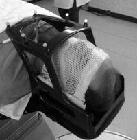
Compensating for patient motion constitutes a challenging as well as important problem for high precision cranial radiotherapy. Common approaches rely on patient immobilization using stereotactic frames, masks or bite bars, or employ X-ray based monitoring and motion compensation such as the 6D skull tracking used by the CyberKnife®. These techniques entail several drawbacks: First, the mask systems are rather inconvenient and also not tolerated by all patients. They further provide no motion monitoring during the treatment - the patient is assumed to be fixed. This is indeed only true up to errors in millimeter range. Second, X-ray based imaging exposes the subject to an unnecessary amount of additional radiation. This exposure also limits the head-tracking speed to about 1 Hz.
Therefore, marker-less optical head-tracking provides a promising alternative, where a laser constantly scans the patient's forehead. This approach requires only light patient fixation and provides a basis for fast real-time monitoring. While promising results have already been achieved using commercial systems such as the Microsoft Kinect®, the accuracy still lags behind the desirable standard of sub-millimeter accuracy. One major drawback of marker-less tracking is given by the lack of point-to-point correspondences. Finding these, for objects which are not strictly rigid, is challenging and prone to registration errors. Point cloud matching algorithms are used which rely, if at all, on a few spatial landmarks. The surface geometry very often introduces further ambiguities which lead to a convergence into local error minima and hence poor robustness.
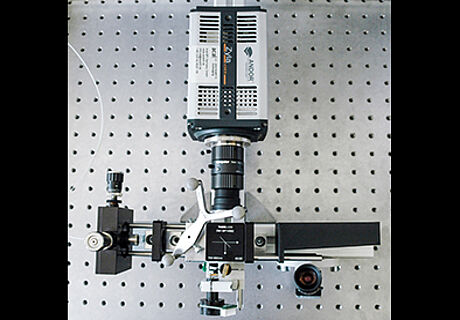
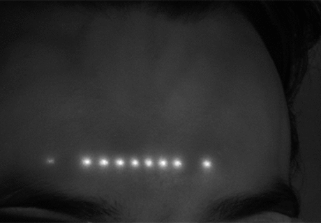
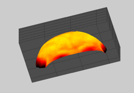
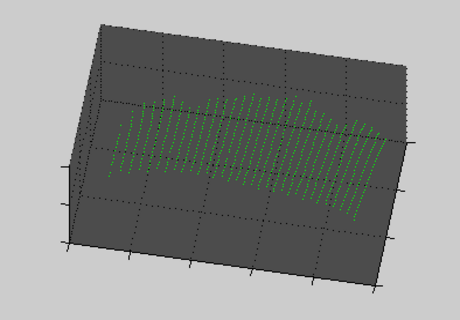
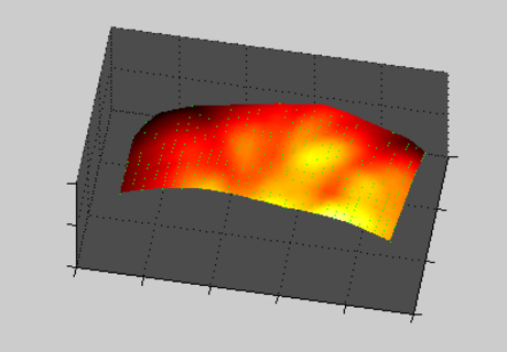
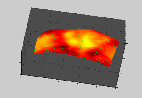
Figure (1) Laser scanning optics, (2) Laser scan of the forehead, (3) MRI segmented tissue ground truth.
By combining expertise from the fields of computer science, electronics, applied mathematics, machine learning and biomedical optics our research group works on laser triangulation, dedicated electronic and optical hardware, as well as enhanced tracking approaches. Specifically the feasibility of optically detecting subcutaneous as well as cutaneous structures is evaluated. The structures serve as supportive landmarks and can be used to localize rigid cranial bone. To uncover these landmarks the developed system extracts optical backscatter features. These are obtained from near-infrared (NIR) laser scans of the forehead. Finally, machine learning algorithms such as Gaussian Processes or Support Vector regression are used to obtain the tissue thickness. In cooperation with Varian Medical, Inc., the world-leading manufacturer for LINACs, we develop a novel prototype for high-accuracy head-tracking in radiotherapy.
Figure (1) Triangulated 3D surface point cloud, (2) feature-labeled point cloud, (3) reconstructed tissue-labeled point cloud.
Publications
2013
Estimating soft tissue thickness from light-tissue interactions a simulation study, Biomedical Optics Express , vol. 4, no. 7, pp. 1176-1187, 2013.
| DOI: | 10.1364/BOE.4.001176 |
| File: | BOE.4.001176 |
Für mehr Präzision in der Tumorbestrahlung, Lübecker Nachrichten , 2013.
| File: | Fuer-mehr-Praezision-in-der-Tumorbestrahlung |
Preliminary Study on Optical Feature Detection for Head Tracking in Radiation Therapy, Chania, Greece , 2013. pp. 1-5.
| DOI: | 10.1109/BIBE.2013.6701632 |
| File: | BIBE.2013.6701632 |
Real time contact-free and non-invasive tracking of the human skull first light and initial validation, San Diego, CA , 2013. pp. 88561G-1 - 88561G-8.
| DOI: | 10.1117/12.2024851 |
| File: | 12.2024851 |
Skin measurements improve motion tracking, 2013.
| File: | 54249 |
Skin thickness estimation for high precision optical head tracking during cranial radiation therapy a simulation study, Atlanta, GA, USA , 2013. pp. S669.
| DOI: | 10.1016/j.ijrobp.2013.06.1775 |
| File: | j.ijrobp.2013.06.1775 |
Time-multiplexed structured light for head tracking, Treuer, Harald, Eds. Cologne, Germany , 2013. pp. 199-202.
2012
Estimation of error sources for optical head tracking in cranial radiation therapy, Düusseldorf, Germany , 2012.
Veröffentlichungen
2013
Estimating soft tissue thickness from light-tissue interactions a simulation study, Biomedical Optics Express , vol. 4, no. 7, pp. 1176-1187, 2013.
| DOI: | 10.1364/BOE.4.001176 |
| File: | BOE.4.001176 |
Für mehr Präzision in der Tumorbestrahlung, Lübecker Nachrichten , 2013.
| File: | Fuer-mehr-Praezision-in-der-Tumorbestrahlung |
Preliminary Study on Optical Feature Detection for Head Tracking in Radiation Therapy, Chania, Greece , 2013. pp. 1-5.
| DOI: | 10.1109/BIBE.2013.6701632 |
| File: | BIBE.2013.6701632 |
Real time contact-free and non-invasive tracking of the human skull first light and initial validation, San Diego, CA , 2013. pp. 88561G-1 - 88561G-8.
| DOI: | 10.1117/12.2024851 |
| File: | 12.2024851 |
Skin measurements improve motion tracking, 2013.
| File: | 54249 |
Skin thickness estimation for high precision optical head tracking during cranial radiation therapy a simulation study, Atlanta, GA, USA , 2013. pp. S669.
| DOI: | 10.1016/j.ijrobp.2013.06.1775 |
| File: | j.ijrobp.2013.06.1775 |
Time-multiplexed structured light for head tracking, Treuer, Harald, Eds. Cologne, Germany , 2013. pp. 199-202.
2012
Estimation of error sources for optical head tracking in cranial radiation therapy, Düusseldorf, Germany , 2012.

- Research
- SonoBox: A Robotic Ultrasound System for Pediatric Forearm Fracture Diagnosis
- Robotics Laboratory (RobLab)
- OLRIM
- MIRANA
- Robotik auf der digitalen Weide
- KRIBL
- Ultrasound Guided Radiation Therapy
- Digitaler Superzwilling: Projekt TWIN-WIN
- - Finished Projects -
- High-Accuracy Head Tracking
- Neurological Modelling
- Modelling of Cardiac Motion
- Motion Compensation in Radiotherapy
- Navigation and Visualisation in Endovascular Aortic Repair (Nav EVAR)
- Autonome Elektrofahrzeuge als urbane Lieferanten
- Goal-based Open ended Autonomous Learning
- Transcranial Electrical Stimulation
- Treatment Planning
- Transcranial Magnetic Stimulation
- Navigation in Liver Surgery
- Stereotactic Micronavigation
- Surgical Microscope
- Interactive C-Arm
- OCT-based Neuro-Imaging
Floris Ernst

Gebäude 64
,
Raum 97
floris.ernst(at)uni-luebeck.de
+49 451 31015200

